CyanoSpectre
Cyanobacteria are fast-emerging as effective chassis for the sustainable synthesis of organic compounds. They are currently being used to produce plastics, proteins, biofuels, and more. Cyanophages are cyanobacteria-infecting viruses that regulate cyanobacterial populations in nature. This makes them a promising vehicle for cyanobacterial engineering. To aid scientists and iGEM teams who work with cyanobacteria, this project aims to create a cyanophage toolkit for their use. Our project will create a “ghost phage” that can recognize and bind marine cyanobacteria. A minimal set of cyanophage genes will be assembled into a phagemid vector that can replicate in E. coli and produce the structural proteins of the phage. To allow our phage to be utilized to immobilize modified cyanobacteria, a biotin-tagged variant of the capsid will be created. Further development of the phage toolkit will enable it to be used as a DNA delivery vehicle in cyanobacterial engineering.
This section is under development.
Confirming A2 Alleles using Luminescence in the Field
Project Inspiration
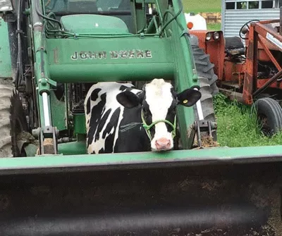
Many small farms in the Oneonta area are struggling to remain solvent in a global, factory-farming marketplace. To remain financially viable, many farmers are turning to niche markets to increase profits and reduce costs by producing specialty products or by selling directly to consumers. In the case of the Dairy industry, one such market it the production of “A2 milk”.
All mammals produce beta-casein, one of the main protein components in milk. All breeds of cow used in the dairy industry produce one of two variants of beta-casein, A1 or A2. Proteolysis of A1 beta-casein can yield a variety of peptides, including beta-casomorphin 7 (BCM7) which belongs to a group of peptides which may have opioid properties (1). Consumption of BCM7 has been linked to a variety of health conditions including type I diabetes, heart disease, and interfering with normal gastrointestinal function (2,3). Furthermore, recent work suggests that many individuals diagnosed with lactose intolerance, may in-fact, be sensitive to A1 beta-casein and BCM7 (4).
While A2 milk is very common in New Zealand, Australia and China, the A2 market in the US is just beginning to expand (5). In the US, most cows are of the A1 lineage, yet, some carry the gene to create A2 milk (6). To create organic A2 milk, it is necessary to identify cows that carry the A2 gene, and to breed them, to create offspring which contain only the A2 gene. While genetic testing can be done to determine a cow’s A status, it is time consuming and expensive, and may take weeks to yield results.
Project goal
The 2020 iGEM team has decided to use synthetic biology to engineer a field-testing system that can quickly and easily provide a dairy farmer with the A status of an animal (A1A1, A1A2 or A2A2). The goal of the project is to make this sensor inexpensive and easy to use, to save farmers time and money, and to promote breeding to create A2 milk cows in the US.
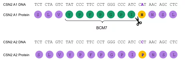
Beta-casein is coded for by the gene CNS2. The two alleles of this gene, A1 and A2, differ by a single nucleotide located in exon 7 (see Figure1) (7). This single nucleotide difference in the DNA sequence creates an amino acid difference in the two versions of the beta-casein protein at amino acid 67. A1 beta-casein contains a Histidine (H) at amino acid 67, while A2 beta-casein contains a Proline (P). The presence of the H creates a cleavage site for proteolysis, and yields the BCM-7 peptide, which does not occur when the P does not. The fact that the two beta-casein alleles differ by only a single nucleotide.
Project Strategy
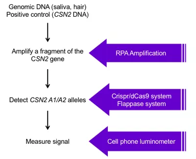
To build the field-deployable testing system, the team identified four critical steps, summarized in Figure 2.
Step 1: Obtain DNA samples for testing.
Ultimately the team will be testing Bovine genomic DNA samples. Such samples may be readily obtained in the field by using a buccal swab to collect check cell DNA, or by taking a small hair sample. In preparation for testing the kit on bovine genomic DNA samples, the team wrote a protocol and received approval from the SUNY Oneonta Institutional Animal Care and Use Committee (IACUC) to collect bovine DNA samples.
Prior to testing the detection systems on genomic DNA samples, the team used synthetic biology to create parts to use as positive controls to validate the assay. To do this, the team searched online databases, identified the region of the CSN2 gene which contains the A1/A2 polymorphism. The team had the A1 and the A2 version of exon 7 synthesized and then cloned the part into a vector for expression in an E.coli chassis. This DNA part can now be easily amplified and used as a positive control during downstream testing.
Step 2: Amplify a fragment of the CSN2 gene
Before the A1/A2 polymorphism can be detected in DNA samples, the region containing the polymorphism must be amplified. Normally, this could be done using PCR, a technique which uses repeated rounds of heating and cooling to copy a piece of DNA. PCR is not a viable technique for a field-deployable kit, traditional DNA amplification techniques will not work, because they rely on expensive equipment for temperature cycling.
Instead, the team elected to use Recombinase Polymerase Amplification (RPA), a technique that can be used to amplify DNA and is performed at one temperature. The team designed and tested primers for use with this technique, and successfully amplified a fragment of the CSN2 gene using the exon 7 positive control constructs as a template.
Step 3: Detect CSN2 A1/A2 Alleles
Because the A1 and A2 version of the CSN2 gene differ by only a single nucleotide, and may be difficult to detect because of this, the team elected to take two different approaches to detect these alleles.
Method 1: Paired Crispr/Cas9 based system.
This detection method is based on a method developed by the Peking 2015 iGEM team, which relies on the use of a guide RNA (gRNA) sequence which binds to, and is used to detect a DNA sequence of interest.
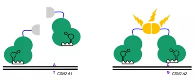
When the DNA of interest is present, the gRNA binds. The gRNA:DNA duplex serves as a docking location for an enzymatically dead Cas9 protein (dCas9), which is used to target other proteins present. In this system, binding of pairs of gRNAs and dCas9 are used to create a light signal, by fusing dCas9 to a fragment of firefly luciferase, which is used to detect binding by emitting visible light (Figure 3).
The iGEM team is currently working on building this system. The students have designed and synthesized 3 pairs of gRNAs to be used to detect the A1 variant, and 3 pairs to detect A2. They are currently working to build the dCas9-luciferase fusion proteins, using a technique called Gibson assembly.
Method 2: Flappase-based system
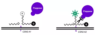
This detection system relies on the use of a Flap Endonuclease (Flappase), an enzyme ubiquitous to life that plays an important role in DNA replication, and repair of DNA breaks (7). In this system, two DNA oligos are designed, to flank and overlap the DNA of interest. The interaction between these two sequences creates a double flap structure which can be recognized and cleaved by the Flappase (8). The longer, flap oligo is cleaved by Flappase if the double flap structure is correct. Cleavage of the flap oligo results in de-quenching of a fluorescent molecule attached to the oligo, which can be detected. A model for how the Flappase system can be used to detect A1 or A2 variants is illustrated in Figure 4.
The team has made good progress on this system. They have cloned the Yeast Flappase gene (Rad 27) and added control elements and purification tags necessary to cause E. coli to produce the large amounts of this enzyme needed to test the system in vitro and are working on purifying the protein and validation. The team has also designed and synthesized the spacer and flap oligos needed to detect A1 and A2 beta-casein, and have created a homology model for Yeast Flappase, based on information available for human Flappase.
Step 4: Measure signal output from the detector system
Both detector systems will yield a signal output (light or fluorescence), which must be measured to determine if the A2 allele is present in a test subject. In a laboratory setting. Light can be measured with a luminometer, and fluorescence intensity can be measured with a fluorimeter. Both pieces of equipment are too large and would be impractical for a field-testing system.
After a literature search, the team discovered that many scientists have been experimenting with building cellphone based portable instrumentation, including light and fluorescence measurement systems (10,11). Based on some designs, the team elected to build a cell phone based luminometer or fluorimeter that can be used in the field to detect signals outputs from the detection systems.
References:
- Jinsmaa and Yoshikawa. 1999. Enzymatic release of neocasomorphin and β-casomorphin from bovine β-casein, Peptides, 20(8): 957-962.
- Nguyen et al. 2015. Formation and Degradation of Beta-casomorphins in Dairy Processing. Critical reviews in food science and nutrition 55(14): 1955-67.
- Jianqin et al. 2016. Effects of milk containing only A2 beta casein versus milk containing both A1 and A2 beta casein proteins on gastrointestinal physiology, symptoms of discomfort, and cognitive behavior of people with self-reported intolerance to traditional cows' milk. Nutrition journal. 5(35): DOI 10.1186/s12937-016-0147-z.
- Pal et al. 2015. Milk Intolerance, Beta-Casein and Lactose. Nutrients. 7(9): 7285-97.
- Sorvino. 2019 Deals With Kroger, Walmart And Costco Fuel U.S. Growth Of A2 Milk Company. Forbes Magazine.
- Pasin. 2017. A2 Milk Facts. California Dairy Research Foundation. May 7, 2020, http://cdrf.org/2017/02/09/a2-milk-facts/.
- Kamiński et al. 2007. Polymorphism of bovine beta-casein and its potential effect on human health. Journal of Applied Genetics 48, 189–198.
- Finger et al. 2012. The wonders of flap endonucleases: structure, function, mechanism and regulation. Sub-cellular biochemistry 62: 301-26. doi:10.1007/978-94-007-4572-8_16.
- Wright et al. 2011. Assays for structure-selective DNA endonucleases. Methods in molecular biology. 745:345-62. doi:10.1007/978-1-61779-129-1_20.
- Friedrichs et al. 2017. SmartFluo: A Method and Affordable Adapter to Measure Chlorophyll a Fluorescence with Smartphones. Sensors 17(4): 678. 25 Mar. 2017, doi:10.3390/s17040678.
- Hossain et al. 2014. Lab-in-a-Phone: Smartphone-Based Portable Fluorometer for pH Measurements of Environmental Water. IEEE Sensors Journal. 15. 10.1109/JSEN.2014.2361651.


